

Compact bone function is essential for keeping the human skeleton strong, stable, and capable of handling the physical demands of everyday life. Also called cortical bone, compact bone forms the dense outer layer of most bones. It provides structural support, protects delicate internal tissues, helps bones resist compression and bending, and creates rigid surfaces that allow muscles to generate effective movement throughout the body.
Although compact bone may look completely solid, it is living tissue with an organized microscopic structure. Blood vessels, nerves, bone cells, collagen fibers, and mineralized matrix work together inside specialized structures called osteons. This design gives compact bone an impressive balance of strength and biological activity. Understanding its structure and location makes it easier to see why this tissue plays such an important role in skeletal health and movement.
What Is Compact Bone?
Compact bone is the dense, hard form of bone tissue that creates the strong external cortex of bones. It is one of the two major structural forms of osseous tissue, the other being spongy or trabecular bone. Its tightly packed organization allows it to withstand significant mechanical forces while preserving the overall shape and stability of the skeleton during standing, walking, running, lifting, and other physical activities.
Despite its solid appearance, compact bone contains microscopic spaces and channels that support living cells. These passages allow blood vessels and nerves to travel through the tissue while providing nutrients to osteocytes. The dense arrangement of mineralized matrix is especially important because it gives bones the rigidity needed to resist bending and compression without making the skeleton completely inflexible or biologically inactive.
What Is the Main Compact Bone Function?
The primary compact bone function is providing strength and structural support to the skeleton. Its dense mineralized tissue helps bones carry body weight and resist the stresses produced by gravity, muscle contraction, and physical movement. This is particularly important in long bones of the arms and legs, where strong cortical walls must repeatedly tolerate compression, tension, twisting, and bending during normal daily activities.
Compact bone also contributes to protection, movement, and mineral storage. Strong cortical tissue shields internal bone structures and supports bones that protect important organs. At the same time, muscles pull against rigid bones to create movement at joints. Bone tissue also acts as an important mineral reservoir, particularly for calcium and phosphate, which are essential for numerous physiological processes throughout the human body.
Compact Bone Structure and Histology
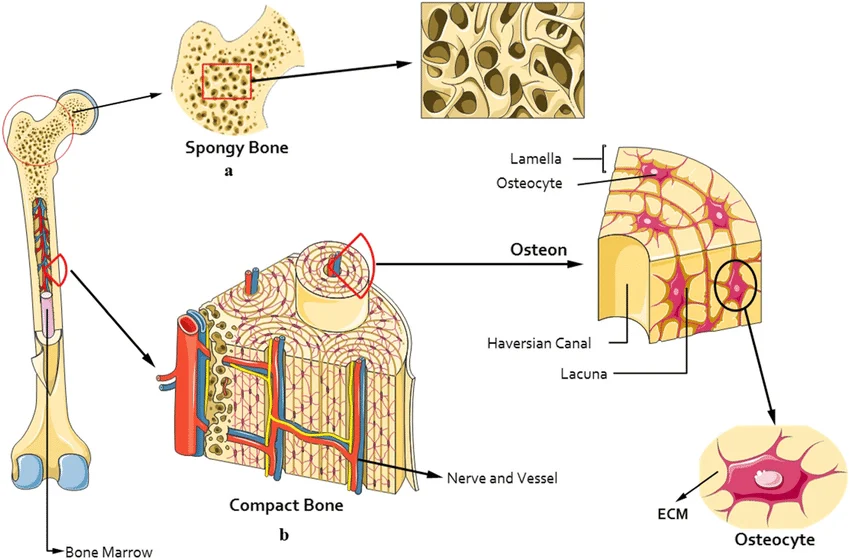
Compact bone has a highly organized microscopic architecture designed to provide both durability and nourishment. Much of the tissue is arranged into cylindrical structures called osteons, also known as Haversian systems. These units contain concentric layers of mineralized matrix surrounding central canals. The arrangement strengthens the bone while creating pathways for blood vessels and nerves that maintain living cells embedded inside the otherwise dense tissue.
The matrix itself contains collagen fibers combined with mineral crystals, especially calcium-containing compounds. Collagen contributes flexibility and resistance to pulling forces, while minerals provide hardness and resistance to compression. This combination prevents healthy bone from behaving like either brittle stone or flexible connective tissue alone. Instead, compact bone achieves a mechanical balance that allows the skeleton to remain strong while tolerating repeated physical stress.
Osteons and Their Role in Compact Bone
Osteons are among the most recognizable structural features of compact bone. Each osteon consists of circular layers called concentric lamellae arranged around a central canal. These cylindrical structures often run roughly parallel to the long axis of bones, helping the tissue withstand forces created during weight-bearing and movement. Their organized arrangement contributes directly to the strength associated with compact bone function.
At the center of an osteon lies the Haversian or central canal, which contains small blood vessels and nerves. Surrounding this canal are layers of mineralized matrix, with mature bone cells positioned within tiny spaces between them. This arrangement ensures that compact bone remains living, responsive tissue rather than an inactive framework. Osteons therefore combine mechanical resistance with access to nutrients and cellular communication.
Lamellae, Lacunae, Osteocytes, and Canaliculi
Concentric lamellae are rings of mineralized bone matrix surrounding the central canal of an osteon. Their layered arrangement helps compact bone withstand stresses from different directions. Collagen fibers within neighboring lamellae may be oriented differently, creating additional resistance to twisting and mechanical strain. This microscopic organization is one reason compact bone can remain strong even when subjected to repetitive movement and substantial loads.
Between the lamellae are small spaces called lacunae that contain mature bone cells known as osteocytes. Tiny channels called canaliculi extend between these spaces, allowing osteocytes to communicate and exchange nutrients and waste products. Because the surrounding matrix is heavily mineralized, this communication network is essential. It enables bone cells to survive, sense changes in mechanical stress, and participate in maintaining healthy skeletal tissue.
Where Is Compact Bone Located?
Compact bone forms the dense outer layer of nearly every bone in the skeleton, although its thickness varies according to location and mechanical demand. It is particularly thick in regions that must withstand substantial stress. Long bones such as the femur, tibia, humerus, radius, and ulna contain prominent layers of compact bone that provide the rigidity required for support and controlled body movement.
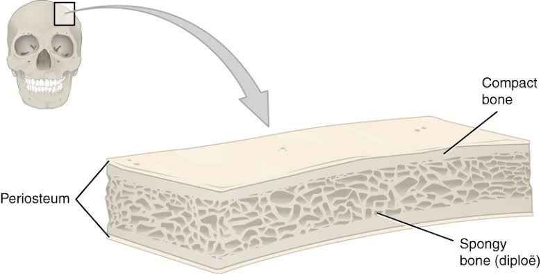
In long bones, compact bone is especially concentrated in the diaphysis, or shaft. This thick cortical wall surrounds the medullary cavity and creates a strong tube-like structure. Compact tissue is also present around spongy bone in the ends of long bones and along the external surfaces of flat, short, and irregular bones. Its distribution reflects the amount and direction of mechanical force experienced in different skeletal regions.
Compact Bone and Weight-Bearing Support
Supporting body weight is a major compact bone function, especially in the lower limbs. During standing and walking, bones such as the femur and tibia experience repeated compressive and bending forces. Their thick cortical walls help distribute these loads efficiently. Without dense compact tissue, long bones would be less capable of supporting the body while maintaining the shape required for stable and coordinated movement.
Compact bone also helps the skeleton tolerate sudden changes in mechanical force. Running, jumping, climbing stairs, and lifting objects can place greater loads on bones than quiet standing. The combination of mineralized matrix and organized osteons allows cortical bone to handle these demands. Its strength therefore supports both everyday activities and more intense movements that temporarily increase stress across joints and long bones.
How Compact Bone Protects the Body
Protection is another essential component of compact bone function. Dense cortical tissue forms strong external surfaces that help shield internal bone structures from physical damage. It also contributes to bones that surround sensitive organs. The skull protects the brain, the rib cage helps protect the heart and lungs, and vertebral structures provide important protection for the spinal cord while supporting the body.
Compact bone also protects internal marrow-containing regions by forming a durable outer shell. This does not mean bones are impossible to fracture, but their dense cortex greatly increases resistance to ordinary mechanical stress. The protective value of compact bone depends on both its mineral content and its organized architecture, allowing it to combine hardness with enough flexibility to tolerate normal physical forces.
Compact Bone Function in Movement
Human movement depends on the close relationship between the skeletal and muscular systems. Muscles connect to bones through tendons and generate force when they contract. Bones then act as rigid levers that move around joints. Compact bone provides much of the stiffness required for these levers to work efficiently, helping convert muscle contraction into controlled actions such as walking, reaching, running, and lifting.
If bones were too flexible, muscle contraction would cause excessive bending rather than efficient movement. Dense cortical tissue helps prevent this problem by maintaining structural rigidity while still allowing bones to absorb normal stresses. The strength of compact bone therefore supports balance, posture, mobility, and athletic performance while working together with muscles, tendons, ligaments, cartilage, and joints throughout the musculoskeletal system.
Compact Bone and Mineral Storage
Bone tissue serves as an important storage site for minerals, particularly calcium and phosphate. These minerals strengthen the bone matrix, but they also perform essential functions elsewhere in the body. Calcium, for example, contributes to muscle contraction, nerve signaling, and many cellular processes. The skeleton therefore functions as both a structural framework and a biologically active mineral reservoir that participates in maintaining internal balance.
Mineral storage adds another dimension to compact bone function because the tissue is constantly involved in remodeling and mineral exchange. Bone can incorporate minerals into its matrix or release them under carefully regulated physiological conditions. This process involves complex hormonal and cellular mechanisms. It demonstrates that compact bone is not simply a permanent shell but a dynamic tissue participating in both mechanical and metabolic functions.
Compact Bone vs. Spongy Bone
Compact bone and spongy bone work together, but their structures are clearly different. Compact bone is dense and usually organized around osteons, while spongy bone consists of a lighter network of trabeculae. Trabecular bone contains numerous open spaces, giving it a porous appearance. This structure helps reduce skeletal weight while allowing forces to be distributed through strategically arranged internal struts.
Their locations also differ. Compact bone forms the strong outer cortex and is especially thick in long-bone shafts, whereas spongy bone is commonly found inside bone ends and within many flat and irregular bones. Neither tissue is unnecessary or superior to the other. Together, they produce bones that combine strength, reduced weight, internal support, and the ability to respond effectively to mechanical loads.
How Compact Bone Responds to Mechanical Stress
Compact bone is living tissue that changes in response to mechanical demands. Everyday activities produce small stresses throughout the skeleton, and bone cells can respond to these forces by influencing tissue maintenance and remodeling. Appropriate mechanical loading helps bones adapt to the demands placed on them, which is one reason physical activity is closely associated with maintaining normal skeletal strength throughout life.
The organization of compact bone also helps distribute stress rather than concentrating it in one small location. Osteons, collagen fibers, and mineralized matrix collectively resist forces created by weight and movement. When microscopic damage occurs, normal remodeling processes can remove and replace affected tissue. This continual renewal helps preserve skeletal integrity as bones repeatedly experience mechanical strain across many years.
Bone Remodeling and Compact Bone Health
Bone remodeling is a continuous biological process involving the removal of older bone and the formation of new tissue. Osteoclasts are specialized cells that break down bone matrix, while osteoblasts produce new matrix that later becomes mineralized. These processes must remain coordinated so the skeleton can repair microscopic damage, adapt to changing demands, and maintain appropriate mineral balance.
Osteocytes also contribute to bone maintenance by sensing changes within their surrounding environment. Through their communication networks, these mature bone cells can help coordinate responses to mechanical stress. Healthy remodeling supports compact bone function by preserving cortical structure and strength. When the balance between bone formation and resorption changes significantly, bone density and resistance to fracture may also change over time.
Compact Bone Through the Lifespan
Compact bone changes throughout life rather than remaining permanently fixed. During childhood and adolescence, skeletal growth increases bone size and modifies cortical structure as the body becomes taller and stronger. Physical activity, nutrition, hormones, genetics, and general health all contribute to the development and maintenance of bone tissue during these important stages of growth.
During adulthood, remodeling continues even after bones stop increasing significantly in length. With aging, changes in bone formation, resorption, and cortical thickness can influence skeletal strength. Maintaining adequate nutrition and appropriate physical activity supports overall bone health, although individual needs differ. The lifelong remodeling process highlights the fact that compact bone remains active, responsive tissue from development through later adulthood.
Why Compact Bone Is Important for Skeletal Health
Healthy compact bone contributes to stability, mobility, posture, and fracture resistance. Because cortical tissue forms the strong external shell of bones, changes in its thickness, density, or microscopic organization can influence overall skeletal performance. Maintaining bone health therefore involves more than simply preserving minerals; the cellular and structural organization of bone tissue must also continue functioning effectively.
The importance of compact bone becomes especially clear when considering how often bones experience stress. Every step, jump, lift, or change in posture transfers forces through the skeleton. Strong cortical tissue allows bones to manage these loads while protecting internal structures. This combination of durability, biological activity, and mechanical efficiency makes compact bone one of the most important structural tissues in the human body.
Conclusion
Compact bone function includes structural support, protection, weight-bearing, movement, resistance to mechanical stress, and mineral storage. Its dense cortical arrangement gives bones the rigidity needed to maintain body shape and support physical activity. At the same time, its internal system of osteons, canals, osteocytes, and blood vessels ensures that compact bone remains living and responsive rather than functioning as inactive structural material.
The relationship between compact bone structure and function demonstrates the remarkable efficiency of skeletal anatomy. Osteons strengthen the tissue, minerals provide hardness, collagen adds resilience, and living cells maintain the matrix through remodeling. Together with spongy bone, compact bone creates a skeleton capable of protecting organs, supporting body weight, enabling movement, storing minerals, and adapting to the changing mechanical demands experienced throughout life.
FAQs
Who is the main structural unit of compact bone?
The osteon, also called the Haversian system, is the main structural unit of compact bone.
Where is compact bone found?
Compact bone forms the dense outer layer of bones and is especially thick in long-bone shafts.
What is the main compact bone function?
Its main functions include strength, support, protection, movement, and resistance to mechanical stress.
Why is compact bone so strong?
Its mineralized matrix, collagen fibers, and organized osteons provide resistance to compression, bending, and twisting.
Is compact bone different from spongy bone?
Yes. Compact bone is dense, while spongy bone contains a lighter network of trabeculae.
Does compact bone contain living cells?
Yes. Osteocytes and other bone cells help maintain and remodel living bone tissue.
You May Also Read: What Is a Microclimate